
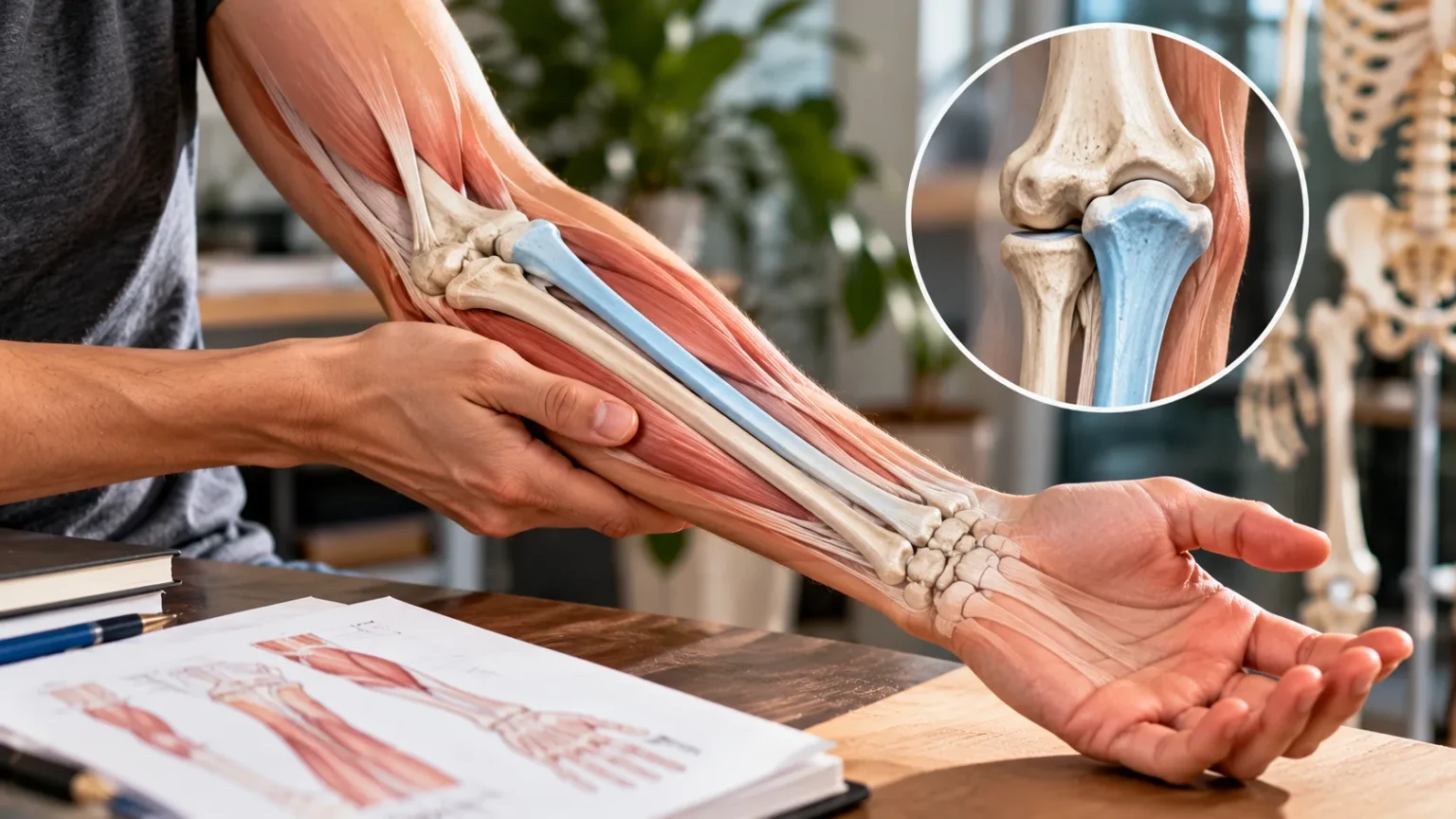

The radius bone is one of the two major bones in the human forearm, playing an essential role in wrist movement, elbow function, and everyday hand activities. Located on the thumb side of the forearm, it works alongside the ulna to support lifting, gripping, and rotating the hand. Understanding its structure helps explain how the upper limb performs movements that often seem effortless.
From turning a doorknob to holding a cup, the radius participates in numerous daily activities. Its unique shape and connections with surrounding joints allow the forearm to rotate while maintaining stability. Although it is a strong long bone, the radius can experience fractures and other injuries, particularly near the wrist.
This guide explores radius bone anatomy, including its location, important anatomical landmarks, joint connections, muscle attachments, and functions. It also explains common radius injuries, symptoms, diagnosis, and general recovery considerations. Whether you are studying human anatomy or simply want to understand your forearm, these details provide a practical introduction.
What Is the Radius Bone?
The radius is a long bone located in the forearm between the elbow and wrist. It lies alongside the ulna and forms important connections with the humerus, wrist bones, and surrounding ligaments. Together, these structures allow the upper limb to perform controlled movements while providing support for muscles and connective tissues.
Unlike the ulna, which contributes substantially to the bony structure of the elbow, the radius has a particularly important relationship with the wrist. Its lower end helps transfer forces between the hand and forearm during activities such as pushing or carrying objects. The radius also rotates around the ulna during forearm movement.
In anatomical terminology, the radius is classified as a long bone because of its elongated structure and specialized regions. It contains a shaft, two ends, and internal bone tissue that provides strength without excessive weight. These characteristics allow the radius to combine structural support with the mobility required for ordinary arm and hand movements.
Where Is the Radius Bone Located?
The radius bone is located on the lateral side of the forearm when the body is in the standard anatomical position. This means it lies on the same side as the thumb when the palms face forward. The ulna occupies the opposite side, closer to the little finger.
The upper end of the radius sits near the elbow, where it connects with the humerus and ulna. Its lower end extends toward the wrist and forms part of the radiocarpal joint. This arrangement allows the radius to participate in movement at both ends of the forearm.
You can identify the thumb-side location of the radius by examining your forearm with your palm facing upward. As you turn your palm downward, the radius crosses over the ulna. This rotational relationship is a defining feature of forearm anatomy and helps explain how the hand changes orientation.
Main Parts of the Radius Bone
The radius consists of three major anatomical regions: the proximal end, shaft, and distal end. Each section has distinctive characteristics that support specific functions. These regions contain articular surfaces, muscle attachment points, and bony landmarks that contribute to the stability and movement of the forearm.
The proximal end is located near the elbow and includes the radial head, neck, and radial tuberosity. The shaft forms the elongated central portion of the bone and provides attachment surfaces for muscles and the interosseous membrane. These structures help connect the radius with surrounding tissues while supporting rotational movement.
The distal end is wider and located near the wrist, where it connects with the carpal bones and distal ulna. It includes important landmarks such as the radial styloid process and ulnar notch. Understanding these anatomical features helps explain common wrist injuries and the radius bone’s role in hand stability.
Anatomy of the Proximal Radius
The proximal radius is the upper portion of the bone closest to the elbow. Its rounded head has a shallow superior surface that articulates with the capitulum of the humerus. The outer surface of the radial head also interacts with the radial notch of the ulna during forearm rotation.
Immediately below the radial head is the radial neck, a narrower region connecting the head to the shaft. This area helps accommodate movement around the elbow but can also be involved in certain injuries. The surrounding annular ligament holds the radial head against the ulna while permitting rotation.
Another important structure is the radial tuberosity, a roughened prominence located below the neck. It serves as the insertion site for the biceps brachii tendon. This attachment allows the biceps muscle to contribute to elbow bending and forearm supination, the movement that turns the palm upward.
Anatomy of the Radius Shaft
The shaft of the radius forms the long central portion connecting the proximal and distal ends. It has a slightly curved shape that contributes to the mechanics of forearm rotation. Anatomically, the shaft contains surfaces and borders that provide attachment points for muscles and connective tissues.
One important feature is the interosseous border, which faces the ulna and provides attachment for the interosseous membrane. This strong connective tissue links the two forearm bones along much of their length. It helps maintain their relationship, provides muscle attachment, and participates in transferring forces across the forearm.
The shaft also supports several muscles involved in wrist, finger, and forearm movements. Its curvature and structural organization allow the radius to rotate relative to the ulna without excessive interference. Damage to the shaft can affect alignment and reduce the normal range of forearm rotation.
Anatomy of the Distal Radius
The distal radius is the broad lower portion of the bone nearest the wrist. It forms a major part of the wrist joint and helps transfer forces from the hand into the forearm. This region is particularly important during weight-bearing activities involving the hands, such as pushing against a surface.
The lower articular surface of the radius connects primarily with the scaphoid and lunate carpal bones. These connections contribute to wrist movements, including flexion, extension, and side-to-side motion. The distal radius also contains the ulnar notch, which articulates with the head of the ulna.
The radial styloid process is a pointed projection on the thumb side of the distal radius. It provides attachment for important wrist-supporting structures and can be felt near the outer wrist. Because the distal radius frequently absorbs force during falls, this region is a common site of fractures.
Radius Bone vs Ulna Bone
The radius and ulna are the two long bones that form the forearm, but they have different anatomical roles. The radius lies on the thumb side, while the ulna lies on the little-finger side in the standard anatomical position. Both contribute to stability and movement between the elbow and wrist.
The ulna has a prominent upper end that forms much of the elbow’s bony structure. In contrast, the radius has a relatively small upper head and a wider lower end that directly supports the wrist joint. These differences reflect how forces and movements are distributed throughout the forearm.
During pronation and supination, the radius moves around the relatively stable ulna. This coordinated action allows the palm to turn downward or upward without requiring the entire upper arm to rotate. Both bones must maintain appropriate alignment for smooth movement and effective hand function.
Joints Connected to the Radius Bone
The radius participates in several important joints that connect the forearm with the elbow and wrist. At the elbow, the radial head articulates with the capitulum of the humerus. This humeroradial articulation contributes to elbow movement and helps transfer forces through the upper limb.
The proximal and distal radioulnar joints allow the radius to rotate relative to the ulna. These joints work together during pronation and supination, making it possible to change the orientation of the palm. Ligaments and surrounding connective tissues provide stability while permitting controlled movement.
At the wrist, the distal radius forms the radiocarpal joint with the proximal carpal bones, mainly the scaphoid and lunate. This connection supports several wrist movements and contributes to load distribution. Healthy joint alignment and surrounding soft tissues are essential for comfortable, coordinated hand activities.
Main Functions of the Radius Bone
One of the primary functions of the radius is supporting movement between the elbow, forearm, and wrist. It allows the hand to rotate into different positions required for daily activities. Without normal radial movement, tasks such as turning keys, opening containers, and using utensils would become considerably more difficult.
The radius also helps transmit forces from the hand toward the upper arm. When you push against a surface or support weight through your palm, forces pass through the wrist into the forearm bones. The radius and surrounding connective tissues distribute these loads while helping maintain structural stability.
Another important function involves providing attachment points for muscles and ligaments. These connections allow muscles to produce controlled movement and help stabilize nearby joints. The radius therefore serves as both a structural support and an active component of the musculoskeletal system.
How the Radius Supports Forearm Rotation
Forearm rotation occurs through two movements called pronation and supination. Pronation turns the palm downward when the elbow is bent, while supination turns it upward. These actions depend on coordinated movement at the proximal and distal radioulnar joints.
During pronation, the radius crosses over the ulna as the forearm rotates. During supination, the radius returns toward a more parallel position alongside the ulna. This arrangement allows substantial changes in hand orientation while preserving the relationship between the elbow and wrist.
Several muscles contribute to these movements, including the pronator teres, pronator quadratus, supinator, and biceps brachii. Their coordinated activity enables both powerful and precise rotational actions. Injuries affecting the radius, joints, or surrounding tissues can interfere with these movements and make ordinary activities uncomfortable.
Muscles Attached to the Radius Bone
Several muscles attach directly to the radius and contribute to upper-limb movement. The biceps brachii inserts into the radial tuberosity, allowing it to assist with elbow flexion and forearm supination. This connection is particularly important when rotating the palm upward against resistance.
The pronator teres attaches to the lateral surface of the radial shaft and helps rotate the forearm into pronation. The pronator quadratus connects the distal radius and ulna, providing another important source of rotational control. Together, these muscles help position the hand for tasks requiring precise movement.
Other muscles associated with the radius include the supinator, brachioradialis, and flexor pollicis longus. These muscles have different origins or insertions along the bone and contribute to forearm, elbow, or thumb function. Their coordinated activity demonstrates how the radius supports multiple movements beyond simple wrist positioning.
Blood Supply and Bone Structure of the Radius
Like other long bones, the radius requires a reliable blood supply to maintain healthy bone tissue. Blood vessels enter through small openings and supply the outer bone, marrow cavity, and surrounding structures. Adequate circulation supports normal bone maintenance and contributes to healing after injuries.
The radius contains a dense outer layer of cortical bone that provides structural strength. Inside certain regions, cancellous bone forms a lighter network of supporting tissue. The internal arrangement varies along the bone according to mechanical demands, particularly near the joints.
Bone tissue constantly undergoes remodeling as specialized cells remove older material and form new tissue. This process helps maintain skeletal strength and supports adaptation to ordinary physical activity. Nutrition, hormones, age, medical conditions, and mechanical loading can influence bone health over time.
Common Injuries Affecting the Radius Bone
Radius injuries may occur during falls, sports activities, road accidents, or other situations involving force through the arm. Fractures are among the most familiar problems affecting this bone. The location and severity of an injury influence symptoms, treatment decisions, and expected recovery.
Distal radius fractures commonly occur near the wrist, particularly when someone falls onto an outstretched hand. Radial head fractures occur closer to the elbow and may affect bending or forearm rotation. Fractures involving the shaft are less common but can interfere significantly with alignment and movement.
Injuries may also involve nearby ligaments, joints, nerves, or connective tissues rather than the bone alone. Pain, swelling, bruising, tenderness, and reduced movement can indicate a problem requiring assessment. Early medical evaluation is important when a fracture or significant joint injury is suspected.
What Is a Distal Radius Fracture?
A distal radius fracture is a break in the lower portion of the radius near the wrist joint. It is one of the more common fractures of the upper limb. Falls onto an outstretched hand are a frequent cause, although the injury can also occur during other accidents.
Symptoms may include wrist pain, swelling, tenderness, bruising, and difficulty moving or using the hand. Some fractures produce visible changes in wrist alignment, while others may be difficult to recognize without imaging. The severity depends on factors such as fracture location, displacement, and involvement of the joint surface.
Treatment depends on the fracture pattern, stability, patient needs, and associated injuries. Some fractures can be managed with immobilization, while others require a procedure or surgery to restore alignment. A qualified healthcare professional determines the appropriate approach after examining the wrist and reviewing diagnostic images.
Radial Head Fractures and Elbow Injuries
A radial head fracture affects the rounded upper end of the radius near the elbow joint. This injury commonly results from force transmitted through the forearm during a fall. It may cause pain around the outer elbow and difficulty rotating the palm upward or downward.
Some radial head fractures involve small cracks or minimal displacement, while others include more complex damage. Symptoms may include swelling, tenderness, restricted elbow movement, and discomfort during everyday tasks. Because the radial head contributes to elbow stability, associated ligament or joint injuries may also require assessment.
Treatment varies according to the fracture’s severity, stability, and the presence of additional injuries. Less severe cases may be managed without surgery, while complex fractures sometimes require surgical treatment. Rehabilitation guidance is important because prolonged stiffness can affect elbow function and forearm rotation.
Symptoms of Radius Bone Problems
Pain is a common symptom of radius injuries, although its exact location depends on the affected region. Distal radius problems usually produce discomfort around the wrist, while proximal injuries may cause pain near the elbow. Shaft injuries can result in tenderness along the forearm.
Swelling, bruising, and difficulty moving the hand or forearm may also occur after an injury. Some individuals notice reduced grip strength or discomfort when rotating the palm. These symptoms can have several causes, so they should not be used alone to identify a specific diagnosis.
Seek urgent medical assessment after a significant injury when there is visible deformity, severe pain, numbness, weakness, or difficulty moving the fingers. Changes in hand color or temperature also require immediate attention. Prompt evaluation helps identify fractures and potentially serious complications affecting circulation or nerve function.
How Doctors Diagnose Radius Bone Injuries
Diagnosing a radius injury usually begins with a medical history and physical examination. A healthcare professional asks how the injury occurred and evaluates the location of pain, swelling, tenderness, and movement limitations. Circulation and nerve function may also be checked to identify associated problems.
X-rays are commonly used to examine suspected radius fractures and assess bone alignment. Images may be taken from several angles to evaluate the affected region properly. These findings help determine whether a fracture is present and whether the nearby joints are involved.
Additional imaging may be recommended for complicated injuries or when ordinary X-rays do not explain persistent symptoms. Computed tomography can provide detailed information about certain complex fractures, while magnetic resonance imaging may help evaluate selected soft-tissue problems. The choice depends on the clinical findings and suspected injury.
Treatment and Recovery of Radius Bone Injuries
Treatment for radius injuries depends on the type of damage, fracture stability, and overall health of the patient. Nonoperative management may involve a splint or cast to protect the affected bone while it heals. Some injuries require surgery when alignment or joint stability cannot be maintained adequately.
Recovery time varies according to fracture severity, treatment, age, and individual healing factors. Bone healing often requires several weeks, but restoring comfortable movement and strength may take longer. Follow-up appointments allow healthcare professionals to assess healing progress and determine when activities can be resumed safely.
Rehabilitation may include guided movement, strengthening, and exercises designed to restore everyday function. These activities should follow the recommendations of the treating clinician, particularly after fractures or surgery. Returning to demanding activities too early can interfere with recovery, so individual instructions are more useful than general timelines.
How to Keep Your Radius Bone Healthy
Maintaining general bone health supports the radius and the rest of the skeletal system. A balanced diet containing sufficient calcium, vitamin D, protein, and other essential nutrients contributes to normal bone maintenance. Individual nutritional needs vary, and supplements are not automatically necessary for everyone.
Regular physical activity can also support muscle strength, coordination, and overall skeletal health. Age-appropriate resistance exercise and other suitable movement activities may help maintain bone strength. The choice of exercise should reflect personal abilities, medical conditions, and any existing injuries rather than following a rigid routine.
Preventing falls and using appropriate protective equipment during relevant activities can reduce the likelihood of certain forearm injuries. People with osteoporosis or other conditions affecting bone strength may benefit from individualized medical guidance. Supporting bone health involves consistent everyday habits rather than relying on one specific exercise or treatment.
Conclusion
The radius bone is an essential component of the forearm that supports wrist movement, elbow function, and rotational control. Its anatomical structure includes the proximal end, shaft, and distal end, each contributing to different mechanical responsibilities. Working with the ulna, muscles, and surrounding joints, the radius helps the upper limb perform precise and coordinated movements.
Understanding radius bone anatomy also makes it easier to recognize why injuries near the wrist or elbow can interfere with everyday activities. Distal radius fractures, radial head injuries, and shaft fractures may affect mobility, strength, and comfort. Proper diagnosis and individualized treatment are important for restoring function and reducing complications.
Maintaining healthy bones, staying physically active, and taking sensible injury-prevention measures can help protect the forearm. Persistent pain or significant injuries should always receive appropriate medical assessment. With an understanding of its anatomy and functions, the importance of the radius in ordinary hand and arm movements becomes much clearer.
Frequently Asked Questions (FAQs)
Where is the radius bone located in the arm?
The radius is located in the forearm on the thumb side when the palm faces forward. It extends from the elbow to the wrist and works alongside the ulna.
What is the main function of the radius bone?
The radius supports wrist movement, helps transmit forces through the forearm, and enables pronation and supination. It also provides attachment points for muscles involved in elbow, forearm, and hand movements.
What is the difference between the radius and ulna?
The radius lies on the thumb side and contributes directly to the wrist joint, while the ulna lies on the little-finger side and forms much of the elbow’s bony structure.
How long does a broken radius bone take to heal?
Many radius fractures show substantial bone healing within approximately six to eight weeks, although timing varies. Complex injuries may require longer, and restoring strength and movement can take several additional months.
Can you move your arm with a fractured radius?
Some people can still move their arm despite a radius fracture, so movement does not rule out injury. Avoid stressing a potentially fractured arm and seek medical assessment for appropriate diagnosis.